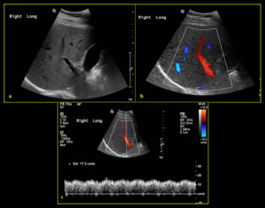
Ultrasound imaging in B-mode, color and spectral Doppler of the
4.6 (77) · $ 11.99 · In stock
Download scientific diagram | Ultrasound imaging in B-mode, color and spectral Doppler of the abdominal organs of the agouti. (a) Ultrasonographic aspects of the urinary vesicle. Note the smooth and echogenic walls with a slight amount of sediment on the interior. (b,d) Color flow and B-mode renal morphology of the right and left kidneys, respectively, showing the usual echotexture and parenchymal echogenicity and preserved corticomedullary limit. (c,e) Pattern of flow of the renal artery, arcuate (arrowhead) and interlobar (arrow) arteries observed with color Doppler. The pulsed Doppler demonstrates well-defined systolic and diastolic peaks. from publication: Abdominal B-mode and Doppler ultrasonography of chemically restrained agouti (Dasyprocta prymnolopha Wagler, 1831) | Agoutis are small-sized wild animals whose body weight can reach up to 4kg, and are found throughout Brazil. They are considered important seed dispersers, especially for big trees and there are species that rely almost exclusively on these animals for their territorial | Doppler Ultrasonography, Doppler Ultrasound and Hemodynamics | ResearchGate, the professional network for scientists.

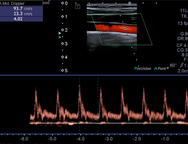
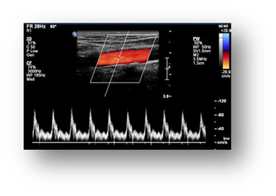
6: Triplex mode image, showing B-mode image, spectral Doppler (bottom

B-mode and color Doppler ultrasonography of normal external jugular vein in donkeys (Equus asinus), BMC Veterinary Research
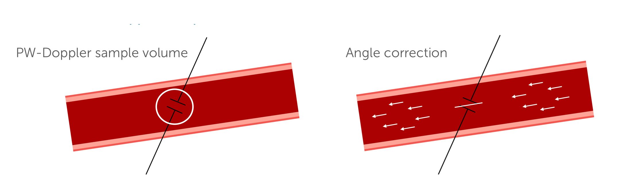
3. Instrumentation and physical principles of carotid (Duplex) ultrasound

presentation-kim-bredahl-doppler-ultrasound.pdf

Caçadores autuados entre 1999 e 2009 nas cidades do entorno do Parque

Doppler Ultrasound - an overview

Caçadores autuados entre 1999 e 2009 nas cidades do entorno do Parque

Flávio ALVES, Laboratory Head, Ph.D, Universidade Federal do Piauí, Teresina, UFPI, Departamento de Morfofisiologia Veterinária

Applied Sciences, Free Full-Text
Spectral Doppler (ultrasound), Radiology Reference Article

B-mode and color Doppler ultrasonography of normal external jugular vein in donkeys (Equus asinus), BMC Veterinary Research
All the Knowledge You Need About Ultrasound Applications and Image Artifacts - Innovatus Imaging
EPOS™

Pigmentation of spleens on subsequent weeks of life, and the

Color Duplex Scanning of the Extracranial Carotid Arteries