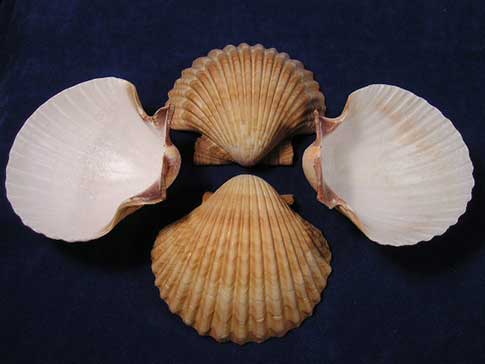
Drawings of the shell of the queen scallop showing (a) the outside
4.9 (111) · $ 6.99 · In stock
Download scientific diagram | Drawings of the shell of the queen scallop showing (a) the outside of the left valve and (b) the inside of the right valve. Illustrations adapted after Lellák & ˆ Cepická (1987). from publication: Fine structural features of the striated adductor muscle of the queen scallop Aequipecten opercularis with respect to age-related changes in intramyocellular components | Age-related changes in the fine structure of the cross-striated (fast) portion of the adductor muscle of the queen scallop, Aequipecten opercularis, were investigated. 14 scallops were sorted into two age groups (for both N = 7) according to shell height: young scallops (≤54 | Pectinidae, Muscle and Myofibrils | ResearchGate, the professional network for scientists.

The Folklore of Shells and Pearls: Scallops, Rhymes and Saints

Electron micrographs showing (a) a tubular aggregate (arrowhead) in a
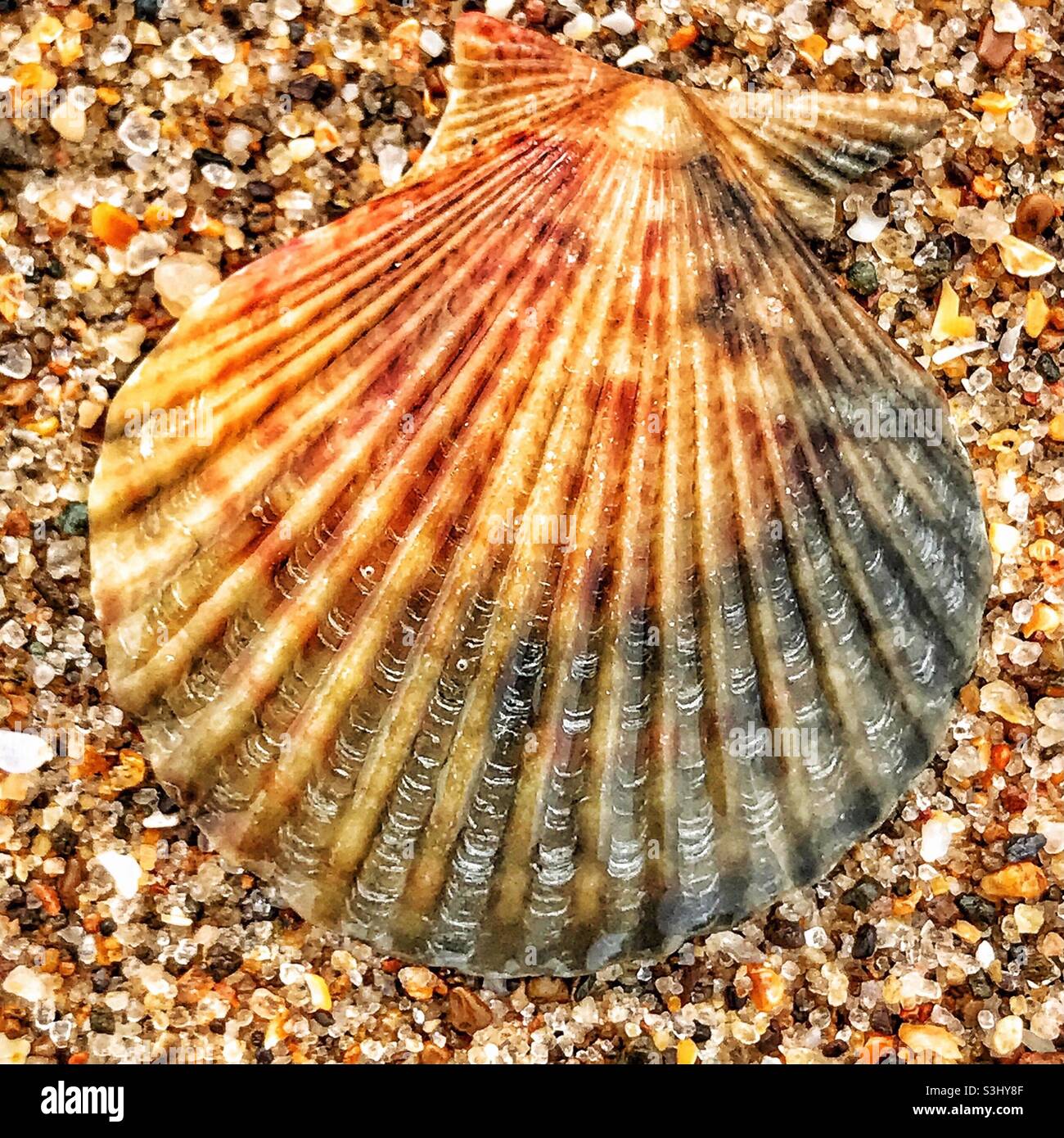
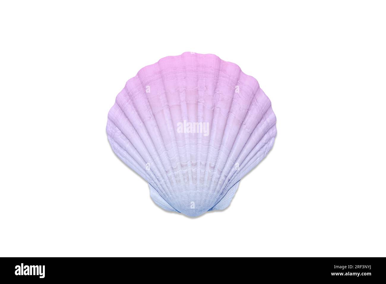
Queen scallop shells hi-res stock photography and images - Alamy
Overview of the pecten body (viewed from the left side with the left

Comparison of three cross-striated teleost muscle fibre types (white

Small amounts of glycogen granules (arrowheads) are visible in the

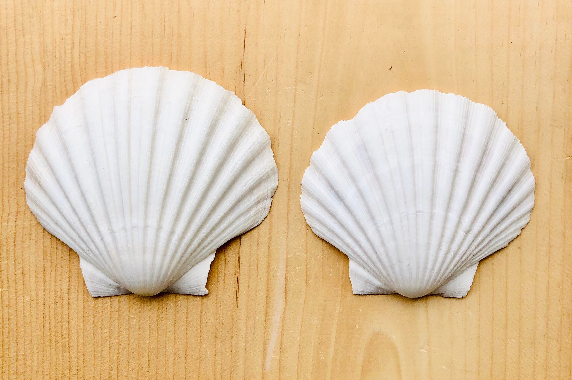
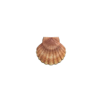
Curved Scallop Seashell, Queen Scallop, Pecten Bovaezelandiae (scientific name), Tupa/Tipa (Maori name), found on…
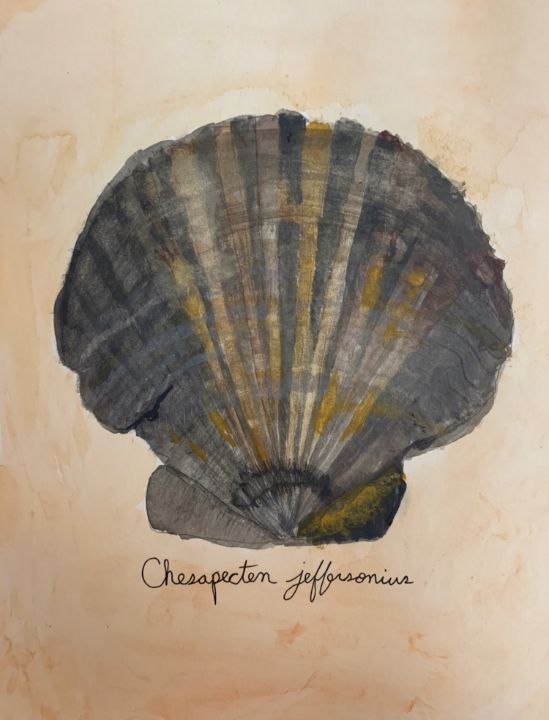
Chesapecten jeffersonius - Andy's Art - Paintings & Prints, Still Life, Shells - ArtPal
Muscle cell nearly filled by the myofibril (Myf) with small amounts of

Isle of Man queen scallops: a shellfish success story
Queen scallop hi-res stock photography and images - Alamy